The Solution: Beams Eye View Tomosynthesis TumoTrack™
Tomosynthesis is a method of producing three-dimensional cross section images in the body by accumulating projection views from a number of view angles and reconstructing the data into tomographic slices. When the X-ray sources positions are set to surround the Therapy beam source, the resulting transverse tomographic images are said to represent the "Beams-Eye-View" and therefore provide a image that displays the cross section of a tumor mass from the view point of the therapy beam. Our simulation have shown that using a tomographic angle of 30 degrees, such slices approach Computer Tomography image quality and are more than adequate for the purpose of tumor tracking.
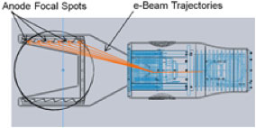
In order to provide BEVT images in realtime, TSS has designed a unique multi-source X-ray tube that uses a small scanning electron beam to move the focal spot from each of 19 focal positions. This tube will be placed just under the Dynamic Multi-Leaf-Collimator (DMLC) of modern linear accelerator based radiotherapy gantries. The projection images will be recorded on the existing Portal Area Detector system which is designed for interlaced kV-MV imaging. The exact tumor contour positions will be determined from the realtime BEVT images and the coordinates provided to the DMLC controller for realtime adjustments of the radiotherapy beam profile. Updates will be provided with a frequency of one per second or faster.
Our TumoTrack™ product is an integrated system consisting of the multi-source tube, integration with the kV-MV portal detector, realtime tomosynthesis reconstruction system, and automatic target recognition software which will be sold on an OEM basis to existing Radiotherapy vendors.
Features
- Realtime tumor of tumors and surrounding soft tissue structures
- Image updates at a rate of 1/sec or faster during actual radiation therapy
- Realtime updates to the treatment plan and adjustment of the DMLC
- No moving parts - focal positions are adjusted electronically with a scanning eBeam
- System may be retrofitted to existing Radiotherapy Gantries
- Provides video sequence record of both the radiotherapy beam and the tumor position during the course of treatment since by saving the Portal Imaging data.
Benefits
- Radiation treatment plans can be more tightly focused on the tumor with less exposure to surrounding tissue
- Higher exposures to the actual tumor volume may be possible increasing the rate of local control and potentially reducing the number of radiation fractions necessary
- The availability of on gantry near-CT quality imaging may improve work flow for the verification of proper patient positioning
- Ultimately improvements in both local control and therapy outcomes can be expected.
Who Would Benefit Using BEVT
Virtually all patients undergoing radiation therapy will benefit from real-time tracking of tumors and surrounding structures can benefit from this product. This product will enable radiation oncologist to develop improved and more effective treatment plans intended to improve the outcomes for patients.
For Radiotherapy providers the addition of a TumoTrack™ system to your gantry will insure that you can provide the latest optimum treatment plans for your patients. In addition, the improvements in positioning, workflow, and portal monitoring may lead to significant reductions in operational and maintenance costs.
For more information on the TSS TumoTrack™ system for Image Guided Radiation Therapy, contact us.
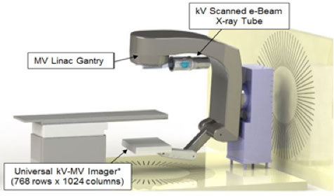
The TumoTrack™ system as mounted on a radiotherapy gantry
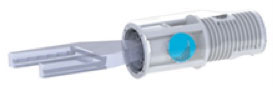
The TSS multi-source X-ray tube
Side view of the TSS multi-source tube